Table Of ContentCurrent Topics in
Microbiology
237 and Immunology
Editors
R.W. Compans, Atlanta/Georgia
M. Cooper, Birmingham/Alabama
J.M. Hogle, Boston/Massachusetts· Y. Ito, Kyoto
H. Koprowski, Philadelphia/Pennsylvania· F. Melchers, Basel
M. Oldstone, La Jolla/California· S. Olsnes, Oslo
M. Potter, Bethesda/Maryland· H. Saedler, Cologne
P.K. Vogt, La Jolla/California· H. Wagner, Munich
Springer
Berlin
Heidelberg
New York
Barcelona
Budapest
Hong Kong
London
Milan
Paris
Singapore
Tokyo
Vascular Growth Factors
and Angiogenesis
Edited by Lena Claesson-Welsh
With 36 Figures and 3 Tables
, Springer
Professor Dr. LENA CLAESSON-WELSH
University of Uppsala
Department of Medical Biochemistry & Microbiology
Biomedical Center
Box 575
S-75l23 Uppsala
Sweden
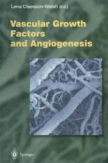
Cover Illustration: Scanning electron microscopy at x 100 ()f' a
micro-vascular corrosion cast of' (/ Wilms' tumor from a 13-month
old boy. Numerous sprouting vessels arc sccn indicating a high
angiogenic actil'ity. The lack of'regulation ()f' angiogcnesis in thc
tumor is apparent through the changes in vcssel-calihre, hiind
cndings and resin leakage. Photo: Dr. Erik SkMdcnberg, Dept. of'
Allatom.\', Biomedical Center, Uppsala, S1t'eden.
Cover Design: design & production GmbH, Heidelberg
ISSN 0070-2 I7X
ISBN-13:978-3-642-64195-4 e-ISBN-13:978-3-642-59953-8
DOl: 10.1007/978-3-642-59953-8
This work is subject to copyright. All rights arc reserved, whether the whole or part of
the material is concerned. specifically the rights of translation, reprinting. rellse of
illustrations. recitation, broadcasting, reproduction on microfilm or in any other way,
and storage in data hanks. Duplication of this puhlication or parts thereof is permilled
only under the provisions of the German Copyright Law of September 9, 1965, in its
current version, and permission for lise must always be obtained from Springer-Verlag.
Violations are liahle for prosecution under the German Copyright Law.
'I', Springer-Verlag Berlin Heidelherg 1999
Softcover reprint of the hardcover 1st edition 1999
Library of Congress Catalog Card Numher 15-12910
The usc of general descriptive names, registered names, trademarks, ctc. in this
publication does not imply, even in the ahsence of a specific statement, that such names
arc exempt from the relevant protective laws and regulations and therefore free for
general usc.
Product liability: The puhlishers cannot guarantee the accuracy of any information
about dosage and application contained in this book. In every individual case the user
must check such information by consulting othcr relevant literaturc.
Typeselling: Scientific Publishing Services (P) Ltd, Madras
Production Editor: Angelique Gcouta
SPIN: 10575594 27J3020 - 543 2 I 0 - Printed on acid-free paper
Preface
Currently, the cellular and molecular mechanisms governing the
development and regulation of the vasculature are studied in
tensely and the field is rapidly progressing. Recently, novel
growth factors and growth factor receptors specifically acting on
endothelial cells have been discovered. Through these factors,
communication networks are established between endothelial
cells, the basement membrane and the pericytes; this interplay is
critical for the regulated development and maintenance of the
vasculature. The awareness that deregulated angiogenesis con
tributes to the progression of a number of diseases, such as cancer
and inflammatory diseases, has clearly spurred the field to move
forward. The focus of this book is on two important classes of
endothelial cell specific growth factors, the vascular endothelial
growth factor (VEGF) family, and the angiopoietins, and on
their mechanisms of action. The reader will find up-to-date, fo
cused reviews, which give the current picture, and indicate future
directions.
I would like to honor Dr. Judah Folkman for his important
contributions to the establishment of the field and thank him for
his support.
Lena Claesson-Welsh
List of Contents
N. FERRARA
Vascular Endothelial Growth Factor: Molecular
and Biological Aspects ......................... .
M. G. PERSICO, V. VINCENTI and T. DIPALMA
Structure, Expression and Receptor-Binding Properties
of Placenta Growth Factor (PIGF) . . . . . . . . . . . . . . . .. 31
U. ERIKSSON and K. ALiTALO
Structure, Expression and Receptor-Binding Properties
of Novel Vascular Endothelial Growth Factors. . . . . . . .. 41
M. SHIBUYA, N. ITo and L. CLAESSON-WELSH
Structure and Function of Vascular Endothelial
Growth Factor Receptor-I and -2 . . . . . . . . . . . . . . . . .. 59
J. TAIPALE, T. MAKINEN, E. ARIGHI, E. KUKK,
M. KARKKAINEN and K. ALiTALO
Vascular Endothelial Growth Factor Receptor-3. . . . . . .. 85
H. F. DVORAK, J. A. NAGY, D. FENG,
L. F. BROWN and A. M. DVORAK
Vascular Permeability Factor/Vascular Endothelial
Growth Factor and the Significance of Microvascular
Hyperpermeability in Angiogenesis . . . . . . . . . . . . . . . .. 97
P. CARMELIET and D. COLLEN
Role of Vascular Endothelial Growth Factor
and Vascular Endothelial Growth Factor Receptors
in Vascular Development ........................ 133
J. PARTANEN and D. J. DUMONT
Functions of Tie I and Tie2 Receptor Tyrosine Kinases
in Vascular Development . . . . . . . . . . . . . . . . . . . . . . .. 159
S. DAVIS and G. D. YANCOPOULOS
The Angiopoietins: Yin and Yang in Angiogenesis 173
Subject Index. . . . . . . . . . . . . . . . . . . . . . . . . . . . . . . .. 187
List of Contributors
(Their addresses can be found at the beginning of their respective chapters.)
ALiTALO, K. 41,85 FERRARA, N.
ARIGHI, E. 85 ITo, N. 59
BROWN, L.F. 97 KARKKAINEN, M. 85
CARMELlET, P. 133 KUKK, E. 85
CLAESSON-WELSH, L. 59 MAKINEN, T. 85
COLLEN, D. 133 NAGY, J.A. 97
DAVIS, S. 173 PARTANEN, J. 159
DIPALMA, T. 31 PERSICO, M.G. 31
DUMONT,D.J. 159 SHIBUYA, M. 59
DVORAK, A.M. 97 TAIPALE, J. 85
DVORAK, H.F. 97 VINCENTI, V. 31
ERIKSSON, U. 41 Y ANCOPOULOS, G.D. 173
FENG. D. 97
Vascular Endothelial Growth Factor:
Molecular and Biological Aspects
N. FERRARA
Introduction
2 Biological Activities of Vascular Endothelial Growth Factor 2
3 Organization of the VEGF Gene and Characteristics of the VEGF Proteins. 4
4 Regulation of VEGF Gene Expression 6
4.1 Oxygen Tension 6
4.2 Cytokines. 6
4.3 Differentiation and Transformation. 7
5 The VEGF Receptors 8
6 The VEGFR-I and VEGFR-2 Tyrosine Kinases. 8
6.1 Binding Characteristics. 8
6.2 Signal Transduction .. 9
6.3 Regulation II
7 Role of VEGF and its Receptors in Physiological Angiogenesis. II
7.1 Distribution ofVEGFR-1 and VEGFR-2 mRNA II
7.2 The VEGFR-I. VEGFR-2 and VEGF Gene Knockouts in Mice 12
8 Role of VEGF in Corpus Luteum Angiogenesis 13
9 Role of VEGF in Pathological Angiogenesis. 14
9.1 Tumor Angiogenesis .. 14
9.2 Angiogenesis Associated with Other Pathological Conditions. 16
10 VEGF and Therapeutic Angiogenesis. 18
II Conclusions. 20
References. 21
1 Introduction
The development of a vascular supply is a fundamental requirement for organ
development and differentiation during embryogenesis as well as for wound healing
and reproductive functions in the adult (FOLKMAN 1995). Angiogenesis is also
implicated in the pathogenesis of a variety of disorders: proliferative retinopathies,
Department of Cardiovascular Research. Genentech, Inc., 460 Point San Bruno Boulevard, South San
Francisco, CA 94080, USA
2 N. Ferrara
age-related macular degeneration, tumors, rheumatoid arthritis and psoriasis
(FOLKMAN 1995; GARNER 1994).
The search for positive regulators of angiogenesis has yielded several candi
dates, including fibroblast growth factors a and b (aFGF, bFGF), transforming
growth factors alpha and beta (TGF-ex, TGF-P), hepatocyte growth factor (HGF),
tumor necrosis factor alpha (TNF-ex), angiogenin, interleukin-8 (IL-8), etc.
(FOLKMAN and SHING 1992; RISAU 1997). However, in spite of extensive research,
there is still significant debate as to their role as endogenous mediators of angio
genesis. The negative regulators identified so far include thrombospondin (GOOD
et al. 1990; DIPIETRO 1997), the 16-kilodalton N-terminal fragment of prolactin
(FERRARA et al. 1991), angiostatin (O'REILLY et al. 1994) and endostatin (O'REILLY
et al. 1997).
This chapter discusses the molecular and biological properties of the vascular
endothelial growth factor (VEGF) proteins. Over the last few years, several addi
tional members of the VEGF gene family have been identified, including VEGF-B,
VEGF-C, Placenta growth factor (PIG F) and VEGF-D. This chapter focuses pri
marily on VEGF, also referred to as "VEGF-A". For a description of the other
members of the family, the reader is referred to the appropriate chapters in this
book. Work done by several laboratories over the last few years has elucidated the
pivotal role of VEGF and its receptors in the regulation of normal and abnormal
angiogenesis (FERRARA and DAVIS-SMYTH 1997). The finding that the loss of even a
single VEGF allele results in embryonic lethality points to an irreplaceable role
played by this factor in the development and differentiation of the vascular system
(FERRARA et al. 1996; CARMELIET et al. 1996). Furthermore, VEGF-induced an
giogenesis has been shown to result in a therapeutic effect in animal models of
coronary or limb ischemia and, most recently, in a human patient affected by
critical leg ischemia (FERRARA and DAVIS-SMYTH 1997).
2 Biological Activities of Vascular Endothelial Growth Factor
Vascular endothelial growth factor (VEGF) is a mitogen for vascular endothelial
cells derived from arteries, veins and lymphatics, but is devoid of consistent and
appreciable mitogenic activity for other cell types (FERRARA and DAVIS-SMYTH
1997). VEGF promotes angiogenesis in tri-dimensional in vitro models, inducing
confluent microvascular endothelial cells to invade collagen gels and form capillary
like structures (PEPPER et al. 1992). Also, VEGF induces sprouting from rat aortic
rings embedded in a collagen gel (NICOSIA et al. 1994). VEGF also elicits a pro
nounced angiogenic response in a variety of in vivo models, including the chick
chorioallantoic membrane (LEUNG et al. 1989), the primate iris (TOLENTINO et al.
1996) etc.
VEGF induces expression of the serine proteases urokinase-type and tissue
type plasminogen activators (PA), and also PA inhibitor 1 (PAl-I) in cultured
Vascular Endothelial Growth Factor: Molecular and Biological Aspects 3
bovine microvascular endothelial cells (PEPPER et al. 1991). Moreover, VEGF in
creases expression of the metalloproteinase interstitial collagenase in human um
bilical-vein endothelial cells (HUVEC), but not in dermal fibroblasts (UNEMORI
et al. 1992). Other studies have shown that VEGF promotes expression of the
urokinase receptor (uPAR) in vascular endothelial cells (MANDRIOTA et al. 1995).
Additionally, VEGF stimulates hexose transport in cultured vascular endothelial
cells (PEKALA et al. 1990).
VEGF is known also as vascular permeability factor (VPF), based on its ability
to induce vascular leakage in the guinea-pig skin (DVORAK et al. 1995). DVORAK
and colleagues proposed that an increase in microvascular permeability is a crucial
step in angiogenesis associated with tumors and wounds (DVORAK 1986). Ac
cording to this hypothesis, a major function of VPFjVEGF in the angiogenic
process is the induction of plasma-protein leakage. This effect would result in the
formation of an extravascular fibrin gel, a substrate for endothelial and tumor cell
growth (DVORAK et al. 1987). Recent studies have also suggested that VEGF may
induce fenestrations in endothelial cells (ROBERTS and PALADE 1995, 1997). Topical
administration ofVEGF acutely resulted in the development offenestrations in the
endothelium of small venules and capillaries, even in regions where endothelial cells
are not normally fenestrated, and was associated with increased vascular perme
ability (ROBERTS and PALADE 1995, 1997).
MELDER et al. (1996) have shown that VEGF promotes expression ofVCAM-l
and ICAM-l in endothelial cells. This induction results in the adhesion of activated
natural killer (NK) cells to endothelial cells, mediated by specific interaction of en
dothelial VCAM-l and ICAM-l with CD18 and VLA-4 on the surface ofNK cells.
VEGF has been reported to have certain regulatory effects on blood cells.
CLAUSS et al. (1990) reported that VEGF may promote monocyte chemotaxis,
while BROXMEYER et al. (1995) have shown that VEGF induces colony formation
by mature subsets of granulocyte-macrophage progenitor cells. These findings may
be explained by the common origin of endothelial and hematopoietic cells and the
presence of VEGF receptors in progenitor cells as early as hemangioblasts in blood
islands in the yolk sac. Furthermore, GABRILOVICH et al. (1996) have reported that
VEGF may have an inhibitory effect on the maturation of host professional anti
gen-presenting cells, such as dendritic cells. VEGF was found to inhibit immature
dendritic cells, without having a significant effect on the function of mature cells.
These findings led to the suggestion that VEGF may also facilitate tumor growth by
allowing the tumor to avoid the induction of an immune response (GABRILOVICH
et al. 1996).
VEGF induces vasodilatation in vitro in a dose-dependent fashion (Ku et al.
1993; YANG et al. 1996) and produces transient tachycardia, hypotension and a
decrease in cardiac output when injected intravenously in conscious, instrumented
rats (YANG et al. 1996). Such effects appear to be caused by a decrease in venous
return, mediated primarily by endothelial cell-derived nitric oxide (NO), as assessed
by the requirement for an intact endothelium and the prevention of the effects by
N-methyl-arginine (YANG et al. 1996). Accordingly, VEGF has no direct effect on
contractility or rate in the isolated rat heart in vitro (YANG et al. 1996). These

