Table Of ContentMICROSCOPY HANDBOOKS 48
Electron Energy Loss Spectroscopy
Royal Microscopical Society MICROSCOPY HANDBOOKS
Series Advisors
Angela Kohler (Life Sciences), Alfred Wegener Institut, Notke-Strasse 85,
22607 Hamburg, Germany
Mark Rainforth (Materials Sciences), Department of Engineering Materials,
University of Sheffield, Sheffield S1 3JD, UK
08 Maintaining and Monitoring the Transmission Electron Microscope
09 Qualitative Polarized Light Microscopy
20 The Operation of Transmission and Scanning Electron Microscopes
21 Cryopreparation of Thin Biological Specimens for Electron Microscopy
25 The Role of Microscopy in Semiconductor Failure Analysis
26 Enzyme Histochemistry
30 Food Microscopy: a Manual of Practical Methods, Using Optical
Microscopy
31 Scientific PhotoMACROgraphy
32 Microscopy of Textile Fibres
33 Modern PhotoMICROgraphy
34 Contrast Techniques in Light Microscopy
35 Negative Staining and Cryoelectron Microscopy: the Thin Film
Techniques
36 Lectin Histochemistry
37 Introduction to Immunocytochemistry (2nd Edn)
38 Confocal Laser Scanning Microscopy
39 Introduction to Scanning Transmission Electron Microscopy
40 Fluorescence Microscopy (2nd Edn)
41 Unbiased Stereology: Three-Dimensional Measurement in Microscopy
42 Introduction to Light Microscopy
43 Electron Microscopy in Microbiology
44 Flow Cytometry (2nd Edn)
45 Immunoenzyme Multiple Staining Methods
46 Image Cytometry
47 Electron Diffraction in the Transmission Electron Microscope
48 Electron Energy Loss Spectroscopy
Forthcoming titles
Energy Dispersive X-ray Analysis in the Electron Microscope
Low Vacuum Scanning Electron Microscopy
High Resolution Electron Microscopy
Electron Energy Loss Spectroscopy
Rik Brydson
LEMAS Centre, Department of Materials, School of Process,
Environmental and Materials Engineering, University of Leeds,
Leeds, UK
@
Taylor & Francis
~ Taylor & Francis Group
LONDON AND NEW YORK
© Taylor & Francis, 2001
First published 2001
All rights reserved. No part ofthis book may be reproduced or
transmitted, in any form or by any means, without permission.
A CIP catalogue record for this book is available from the British Library.
ISBN 1 85996 134 7
Published by Taylor & Francis
2 Park Square, Milton Park, Abingdon, Oxon, OX14 4RN
52 Vanderbilt Avenue, New York NY 10017
Transferred to Digital Printing 2006
Production Editor: Paul Barlass.
Typeset by Marksbury Multimedia Ltd, Midsomer Norton, Bath, UK.
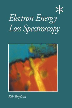
Front cover: RGB processed image obtained from three EFTEM
elemental maps of the interface between a boride-coated fibre and a
titanium alloy matrix in a fibre reinforced metal matrix composite. Red,
titanium; green, boron; blue, gadolinium. Image reproduced with
permission from Brydson R, Hofer F, et al., Micron 1996; 27: 107-120.
Image recorded at FELMI-TUGRAZ by F. Hofer.
Publisher's Note
The publisher has gone to great lengths to ensure the quality of this reprint
but points out that some imperfections in the original may be apparent
Contents
Abbreviations ix
Preface xi
Acknowledgements xii
1. Introduction 1
What is EELS? 1
Interaction of electrons with matter 2
Basics of the TEM 17
Comparison of EELS in TEM with other spectroscopies 20
Conclusions 25
References 25
2. The EEL spectrum 27
The primary transmitted electron signal 27
Historical development 27
Basic components of an EEL spectrum 29
Basic physics 32
Summary of analytical uses 36
References 37
3. EELS instrumentation and experimental aspects 39
The electron spectrometer 39
Coupling a magnetic spectrometer to the microscope 42
Spectral recording 44
Energy-filtered imaging 49
Choice of experimental conditions for EELS 51
Specimen parameters 56
Summary of experimental set-up and data acquisition
procedures for EELS 57
Summary of data correction procedures for EELS 58
References and further reading 58
v
vi Electron Energy Loss Spectroscopy
4. Low loss spectroscopy 59
Quantification of sample thickness 59
Quantitative aspects of low loss data 61
Experimental aspects of EELS low loss measurements 66
Conclusions 67
References and further reading 67
5. Elemental quantification 69
Quantification of EEL spectra 69
Background removal 70
Determination of the ionization cross-section 72
Final quantification step 75
Summary of the quantification procedure 76
Summary of experimental quantification parameters 76
Accuracy and detection sensitivity of EELS quantification
and comparison with EDX in the TEM 77
References and further reading 78
6. Fine structure on inner-shell ionization edges
(ELNESIEXELFS) 79
Origin of edge fine structure 79
Determination of coordinations 84
Determination of valencies 88
Determination of bond lengths 91
Experimental aspects of ELNESIEXELFS measurements 93
Conclusions 95
References and further reading 95
7. EELS imaging 97
Introduction to EELS imaging and energy filtering 97
Summary of energy-filtering techniques 99
Procedure for EFTEM elemental mapping 101
Experimental parameters in EFTEM 104
Correlation of elemental maps 109
General strategy for EFTEM elemental analysis 109
Experimental procedure for EFTEM image acquisition and
processing 110
Comparison of EFTEM and spectrum imaging methods 111
Energy-filtered tomography 112
Conclusions 113
References and further reading 113
Contents vii
8. Advanced EELS techniques in the TEM 115
Orientation dependency in EELS 115
Spatially resolved measurements 118
Electron Compton scattering 125
Reflection mode and surface measurements 126
References and further reading 127
9. Conclusions 129
Further reading 129
Further resources 130
Index 133
Abbreviations
ADC analogue to digital converter
ADF annular dark field
AES Auger electron spectroscopy
ALCHEMI atom location by channelling-enhanced microanalysis
AO atomic orbital
BF bright field
BIS Bremstrahlung isochromat spectroscopy
CCD charge coupled device
CL cathodoluminescence
CTEM conventional TEM
DF dark field
DFT density functional theory
DOS density of states
DQE detector quantum efficiency
ECOSS electron Compton scattering from solids
EDX energy dispersive X-ray
EELS electron energy loss spectroscopy
EFTEM energy-filtered transmission electron microscopy
ELNES electron energy loss near-edge structure
EPMA electron microprobe analyser
ESI electron spectroscopic imaging
EXAFS extended X-ray absorption fine structure
EXELFS extended electron energy loss fine structure
FIB focused ion beam
FIM field ion microscopy
FWHM full width half maximum
GIF Gatan imaging filter
GOS generalized oscillator strength
HAADF high-angle annular dark field
HREELS high resolution electron energy loss spectroscopy
IPS inverse photoemission spectroscopy
IR infrared
JDOS joint density of states
LAMMA laser microprobe mass analysis
LDA local density approximation
MASNMR magic angle spinning nuclear magnetic resonance
MO molecular orbital
ix

