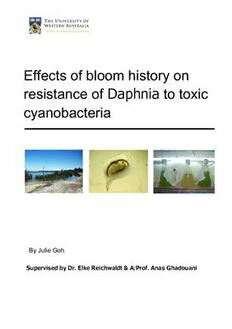
Table Of ContentEffects of bloom history on
resistance of Daphnia to toxic
c yanobacteria
By Julie Goh
Supervised by Dr. Elke Reichwaldt & A/Prof. Anas Ghadouani
Effects of bloom history on resistance of Daphnia to toxic cyanobacteria i
This page has intentionally been left blank
ii Effects of bloom history on resistance of Daphnia to toxic cyanobacteria
The following dissertation has been completed in partial fulfilment of the requirements
of the Bachelor of Engineering (Environmental Systems Engineering) course at the
University of Western Australia.
Effects of bloom history on resistance of Daphnia to toxic cyanobacteria iii
This page has intentionally been left blank
iv Effects of bloom history on resistance of Daphnia to toxic cyanobacteria
Abstract
The common presence of Daphnia within water bodies on an international basis and their significance
in ecosystems establishes this zooplankton genus as a keystone species. The non-selective grazing
habits of Daphnia and the presence of cyanobacterial blooms in such lakes where Daphnia is found,
means there is potential for the accumulation of cyanobacterial toxins in Daphnia and a subsequent
transfer of the toxins to animals higher in the food web for predators such as fish. Additionally,
abundance of Daphnia in water bodies also has a direct effect on the quantity of food sources
available to secondary consumers such as fish. Studying the effects of toxic cyanobacteria on
Daphnia therefore has potential benefits for understanding ecosystems. Although further research is
required, the study may also be able to contribute to determining whether Daphnia is suitable for bio-
manipulation, particularly for the purpose of controlling cyanobacterial blooms in Australia. The aim of
this study was to determine whether Daphnia from lakes with more frequent bloom histories would be
less affected by greater cyanobacterial concentrations in relation to the juvenile growth rate and
survival rate.
Samples of Daphnia were taken from three shallow freshwater lakes in the Perth metropolitan area.
Individuals from each of the lakes were identified as being of the same species using a taxonomic key
and microscopic observation. Laboratory cultures were grown from a single parthenogenetic female
sourced from each respective lake using spring water as growth medium and Desmodesmus as the
food source. In the experiments, cultures from the lakes were tested at four concentrations of toxic
Microcystis aeruginosa: 0%, 20%, 60% and 100%, measured as a fraction of carbon mass of the total
food source. Quantities of food source were such that a concentration of 1mg carbon per Litre was
maintained and consisted of Desmodesmus and toxic M. aeruginosa. Survival rates were measured
by the introduction of neonates born within 3 days of each other into the respective concentrations for
6 days and were monitored daily. Juvenile growth rate was determined by measuring the mass of
neonates after three days in the respective treatment, and comparing it to the initial masses.
The results showed an increase in survival rate with higher M. aeruginosa concentration for Lake
Yangebup, while Lake Monger and Jackadder Lake showed a decrease in survival rate for the same
conditions. These contrasting responses to M. aeruginosa were accounted for by the high microcystin
concentration in the bloom history of Lake Yangebup compared to Lake Monger and Jackadder Lake.
The decrease in juvenile growth rate by mass with increasing toxic M. aeruginosa concentration is
indicative of the inhibition of growth resulting from cyanobacteria content despite an insignificant p-
value.
Effects of bloom history on resistance of Daphnia to toxic cyanobacteria v
Acknowledgements
First and foremost, I would like to thank is Dr Elke Reichwaldt for her guidance, time and
continuous support over the past year. Her enthusiasm to the project and never-ending patience
and feedback has greatly aided completion of the following dissertation. I would also like to thank
Associate Professor Anas Ghadouani for his insight and support during times of need. Many
thanks also to Shian Min Liau for her guidance and patience; advancement of the project was
significantly aided by her help.
Regards to Dianne Krikke for facilitating the needs of the project in the lab; as well as Nicola
Kingdon, Liah Coggins, Haihong Song and Som Cit Si Nang for their constructive criticism during
the review of project work.
Acknowledgements to Darryl Roberts, Michael Smirk, Jemima, Grzegorz Skrzypek and Douglas
Ford for their assistance in obtaining use of equipment used in the experimental component of
the project.
A big thank you to my peers both within and outside of the SESE for their advice, friendship,
support, company and entertainment provided over the time; the plethora of hours spent working
on this dissertation would have led to insanity without their presence.
And lastly but most importantly, many thanks to my family for their everlasting patience, support
and understanding.
vi Effects of bloom history on resistance of Daphnia to toxic cyanobacteria
Contents
Abstract.....................................................................................................................................................v
Acknowledgements ................................................................................................................................. vi
List of Figures .......................................................................................................................................... 1
List of Tables ............................................................................................................................................x
List of Acronyms .......................................................................................................................................x
1. Introduction ...................................................................................................................................... 1
2. Background...................................................................................................................................... 3
2.1. Toxic Cyanobacteria and microcystin ...................................................................................... 3
2.1.1. Microcystin ....................................................................................................................... 3
2.1.2. Bloom formation............................................................................................................... 4
2.2. Cyanobacteria in the ecosystem ............................................................................................. 5
2.3. Daphnia and Ecosystems ........................................................................................................ 6
2.4. Daphnia ................................................................................................................................... 7
2.4.1. Distribution ....................................................................................................................... 8
2.4.2. Daphnia as a Bioindicator ................................................................................................ 9
2.4.3. Risks to Daphnia: Field vs. Lab ..................................................................................... 10
2.5. Toxicology Studies ................................................................................................................ 11
2.6. Study Sites............................................................................................................................. 13
3. Methods ......................................................................................................................................... 14
3.1. Aims ....................................................................................................................................... 14
3.2. Sites ....................................................................................................................................... 15
3.2.1. Field work ..................................................................................................................... 16
3.3. Experimental Design ............................................................................................................. 17
3.4. Cultures ................................................................................................................................. 18
3.4.1. Desmodesmus ............................................................................................................... 18
3.4.2. Microcystis aeruginosa .................................................................................................. 19
3.4.3. Food Source Preparations ............................................................................................. 20
3.5. Daphnia Cultures ................................................................................................................... 22
3.6. Survival Tests ........................................................................................................................ 24
3.7. Juvenile Growth Rate Tests .................................................................................................. 25
4. Results ........................................................................................................................................... 27
4.1. Survival Rate ......................................................................................................................... 27
Effects of bloom history on resistance of Daphnia to toxic cyanobacteria vii
4.1.1. Comparisons between Concentrations ......................................................................... 27
4.1.2. Comparison between Lakes .......................................................................................... 30
4.2. Juvenile Growth Rate ............................................................................................................ 32
5. Discussion ..................................................................................................................................... 33
5.1. Survival Rate ......................................................................................................................... 33
5.1.1. Comparison between Concentrations ........................................................................... 33
5.1.2. Comparison between Lakes .......................................................................................... 35
5.2. Juvenile Growth Rate ............................................................................................................ 36
5.3. Laboratory/ Equipment Faults ............................................................................................... 37
6. Future Recommendations ............................................................................................................. 38
6.1. Non-toxic M. aeruginosa Treatment ...................................................................................... 38
6.2. Bloom History Variation ......................................................................................................... 38
6.3. Experimental Design ............................................................................................................. 38
6.4. Calibration Curve Technique ................................................................................................. 39
7. Conclusion ..................................................................................................................................... 40
8. References .................................................................................................................................... 41
Appendix A: Water Quality Analysis .................................................................................................. 43
Appendix B: WC Medium Preparation ............................................................................................... 44
Appendix C: SPE Protocol ................................................................................................................. 45
Appendix D: HPLC Protocol ............................................................................................................... 46
Appendix E: HPLC Analysis ............................................................................................................... 48
Appendix F: Food Source Calculation ............................................................................................... 49
Appendix G: Survival Experiment Form ......................................................................................... 51
Appendix H: FluoroProbe Analysis of Survival Experiment Medium ................................................. 52
Appendix I: Juvenile Growth Rate Data ............................................................................................ 53
Appendix J: Survival Data Graphs .................................................................................................... 54
viii Effects of bloom history on resistance of Daphnia to toxic cyanobacteria
List of Figures
Figure 1.1. Schematic summary of factors and their influence on microcystin (MC) production, adapted from
Zurawell et al. (2004). .................................................................................................................................. 3
Figure 1.2. Photo of Lake Yangebup and the accumulation of cyanobacteria along the shore. Photo taken by J
Goh. .............................................................................................................................................................. 4
Figure 1.3. Web of influence of cyanobacteria on ecosystems, adapted from Zurawell et al. (2005). ................... 5
Figure 1.4. Schematic illustration of a daphnid, adapted from (Peters & Bernardi 1987). ..................................... 7
Figure 1.5. Map showing distribution of Daphnia where closed circles denote locations at where Daphnia were
found and open circles denote locations where Daphnia was not found (Benzie 1988).............................. 8
Figure 1.6. Typical Daphnia life cycle from neonate to release of first clutch. Photos taken by J. Goh. ................. 9
Figure 1.7. Graphs of cyanobacteria and microcystin concentrations for each site (Nang et. al., submitted). .... 13
Figure 2.1. Map of Perth region showing sampled lakes and aerial photos of Jackadder Lake (top), Lake Monger
(middle) and Lake Yangebup (bottom). ..................................................................................................... 15
Figure 2.2. Photos of samples taken from Jackadder Lake (top), Lake Yangebup (middle) and Lake Monger
(bottom). .................................................................................................................................................... 16
Figure 2.3. Photo (left) of Desmodesmus culture in laboratory conditions and illustration (right) of culturing
equipment setup. ....................................................................................................................................... 18
Figure 2.4. Chromatogram from HPLC-PDA analysis of one from three analysed filters. ..................................... 19
Figure 2.5. Analysed peaks and characteristic shape of MC-LR peaks. ................................................................. 20
Figure 2.6. Calibration curve relating absorption to carbon content for Desmodesmus sp. (established by Liau
2010). ......................................................................................................................................................... 20
Figure 2.7. Calibration curve relating absorption to carbon content for toxic M. aeruginosa (established by Liau
2010). ......................................................................................................................................................... 21
Figure 2.8. Photos of female adults taken with 4x magnification of sample representative individuals from i)
Jackadder Lake ii) Lake Monger and iii) Lake Yangebup for species comparison. Photo taken by J. Goh. 23
Figure 2.9. Photo of experimental setup for survival testing of Jackadder Lake individuals. Photo taken by J. Goh.
................................................................................................................................................................... 24
Figure 2.10. Schematic diagram of the experimental design for testing of the juvenile growth rate of Lake
Yangebup individuals. ................................................................................................................................ 25
Figure 3.1. Limit survival estimates produced using JMP IN for Lake Yangebup. ................................................. 27
Figure 3.2. Limit survival estimates produced using JMP IN for Lake Monger. ..................................................... 27
Figure 3.3. Limit survival estimates produced using JMP IN for Jackadder Lake. ................................................. 28
Figure 3.4. Comparison of survival curves between lakes for 0% treatments (fed only Desmodesmus as the food
source). ...................................................................................................................................................... 30
Figure 3.5. Comparison of survival curves between lakes for 100% treatments (fed only M. aeruginosa as the
food source). .............................................................................................................................................. 30
Figure 3.6. Plot of mean values of juvenile growth rate and corresponding error bars. ....................................... 32
Figure 4.1. Graphs of bloom history for the three lakes from which Daphnia individuals were taken, adapted
from Nang et al. (submitted). .................................................................................................................... 33
Effects of bloom history on resistance of Daphnia to toxic cyanobacteria ix
Figure A. 1. Survival data plots comparing lakes for a) 100% treatment; b) 60% treatment; c) 20% treatment and
d) 0%. ......................................................................................................................................................... 54
Figure A. 2. Survival data plots for a) Lake Yangebup (top-left); b) Lake Monger (top right); c) Jackadder Lake
and d) raw survival data for all treatments. .............................................................................................. 55
List of Tables
Table 2.1. Coding system used for labelling replicates. ........................................................................................ 17
Table 3.1. Corresponding statistical information from survival analysis of treatments for each lake produced
from JMP IN. .................................................................................................................................................... 28
Table 3.2. Matrix showing p-values of statistical analyses used to identify significant differences in
concentration groups for each lake. ................................................................................................................ 29
Table 3.3. Statistics from log rank tests for comparison of 0% and 100% M. aeruginosa treatments between
lakes. ................................................................................................................................................................ 31
Table 3.4. Statistics from one-way ANOVA analysis performed on juvenile growth rate data. ............................ 32
Table A. 1. List of parameters used for FluoroProbe analysis. .............................................................................. 43
Table A. 2. Field measurements and phytoplankton composition of lake water. ................................................. 43
Table A. 3. Calculation of food source composition for 23rd of April 2010. .......................................................... 50
Table A. 4. Data and calculation of juvenile growth rates. ................................................................................... 53
List of Acronyms
ERL – Environmental Research Laboratory
FNAS – Faculty of Natural and Agricultural Sciences
HPLC-PDA – High Performance Liquid Chromatography with Photo-Diode Array
MC-LR – microcystin containing leucine and arginine
M. aeruginosa – Microcystis aeruginosa
POC – Particulate Organic Carbon
SEE – School of Earth and Environment
SESE – School of Environmental Systems Engineering
SPE – Solid Phase Extraction
UWA – The University of Western Australi
x Effects of bloom history on resistance of Daphnia to toxic cyanobacteria

