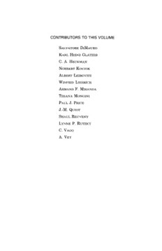
Table Of ContentCONTRIBUTORS TO THIS VOLUME
SALVATORE DIMAURO
KARL HEINZ GLÄTZER
C. A. HECKMAN
NORBERT KOCIOK
ALBERT LEIBOVITZ
WlNFRID LlEBRICH
ARMAND F. MIRANDA
TlZIANA MONGINI
PAUL J. PRICE
J.-M. QUIOT
SHAUL REUVENY
LYNNE P. RUTZKY
C. VAGO
A. VEY
Advances in
CELL CULTURE
Edited by
KARL MARAMOROSCH
Robert L. Starkey Professor of Microbiology
Waksman Institute of Microbiology
Rutgers University
New Brunswick, New Jersey
VOLUME 4
1985
ACADEMIC PRESS, INC.
(Harcourt Brace Jovanovich, Publishers)
Orlando San Diego New York London
Toronto Montreal Sydney Tokyo
COPYRIGHT © 1985, BY ACADEMIC PRESS, INC.
ALL RIGHTS RESERVED.
NO PART OF THIS PUBLICATION MAY BE REPRODUCED OR
TRANSMITTED IN ANY FORM OR BY ANY MEANS, ELECTRONIC
OR MECHANICAL, INCLUDING PHOTOCOPY, RECORDING, OR
ANY INFORMATION STORAGE AND RETRIEVAL SYSTEM, WITHOUT
PERMISSION IN WRITING FROM THE PUBLISHER.
ACADEMIC PRESS, INC.
Orlando, Florida 32887
United Kingdom Edition published by
ACADEMIC PRESS INC. (LONDON) LTD.
24-28 Oval Road, London NW1 7DX
ISSN Ο275-6358
ISBN 0-12-007904-6
PRINTED IN THE UNITED STATES OF AMERICA
85 86 87 88 9 8 7 6 5 4 3 2 1
CONTRIBUTORS TO VOLUME 4
Numbers in parentheses indicate the pages on which the authors' contributions begin.
SALVATORE DIMAURO (1), Department of Neurology, Columbia Univer-
sity, College of Physicians and Surgeons, and the H. Houston Mer-
ritt Clinical Research Center for Muscular Dystrophy and Related
Diseases, New York, New York 10032
KARL HEINZ GLÄTZER (179), Institut für Genetik, Universität Düsseldorf,
D-4000 Düsseldorf, Federal Republic of Germany
C. A. HECKMAN (85), Department of Biological Sciences, Bowling Green
State University, Bowling Green, Ohio 43403
NORBERT KOCIOK (179), Institut für Genetik, Universität Düsseldorf,
D-4000 Düsseldorf, Federal Republic of Germany
ALBERT LEIBOVITZ (249), Arizona Cancer Center, University of Arizona
College of Medicine, Tucson, Arizona 85724
WINFRID LIEBRICH (179), Institut für Genetik, Universität Düsseldorf,
D-4000 Düsseldorf, Federal Republic of Germany
ARMAND F. MIRANDA (1), Department of Pathology, Columbia Univer-
sity, College of Physicians and Surgeons, and the H. Houston Mer-
ritt Clinical Research Center for Muscular Dystrophy and Related
Diseases, New York, New York 10032
TIZIANA MONGINI (1), Department of Neurology, Columbia University,
College of Physicians and Surgeons, and the H. Houston Merritt
Clinical Research Center for Muscular Dystrophy and Related Dis-
eases, New York, New York 10032
PAUL J. PRICE (157), Hybridoma Sciences, Inc., Atlanta, Georgia 30084
J.-M. QUIOT (199), Centre de Recherches de Pathologie Comparée, INRA,
CNRS, USTL, 30380 Saint-Christol, France
SHAUL REUVENY l (213), Department of Biotechnology, Israel Institute for
Biological Research, Ness-Ziona 70450, Israel
LYNNE P. RUTZKY (47), Department of Surgery, The University of Texas
Medical School, Houston, Texas 77030
C. VAGO (199), Centre de Recherches de Pathologie Comparée, INRA,
CNRS, USTL, 30380 Saint-Christol, France
A. VEY (199), Centre de Recherches de Pathologie Comparée, INRA,
CNRS, USTL, 30380 Saint-Christol, France
1Present address: New Brunswick Scientific Co., Inc., 44 Talmadge Rd., Edison, New
Jersey 08818.
IX
PREFACE
Volume 4 of Advances in Cell Culture continues the wide coverage of
this serial publication. Chapters are devoted to basic aspects such as
cell shape and growth control in vitro, morphological and cytochemical
changes occurring during muscle culture of human myopathies, and
the biology of cultured human colon tumor cells. Among the important
practical topics are the structure and application of microcarriers and
hybridoma technology. One chapter is devoted to the establishment of
cell lines from human solid tumors. The use of invertebrate organ
culture for the study of spermiogenesis and the study of mycotoxin
action on invertebrate cells are presented in two chapters. Readers will
find stimulating ideas, new concepts, and fresh approaches to problems
in the current contributions. Facets of cell culture that may have im-
mediate, as well as long-range economic potential, are presented. The
contributions reflect the thinking and accomplishments of those who
are in the forefront of the broad field of cell culture today. The depth
and sophistication of the articles indicate current strength in the di-
verse areas of in vitro research.
In this volume, a biographical sketch has been devoted to George
Gey, who had a profound impact on cell culture and on numerous
scientists who became inspired by him.
KARL MARAMOROSCH
XI
GEORGE GEY
Xll
GEORGE GEY
Xll
DREAMS OF CELLULAR PHYSIOLOGY IN TISSUE CULTURE:
GEORGE GEY S BEQUEST
In 1923, Dr. John L. Yates, a graduate of The Johns Hopkins Univer-
sity School of Medicine and Chief of Surgery at Columbia Hospital in
Milwaukee, Wisconsin, and whose interest was in cancer of the breast,
wrote to Dr. Warren Lewis of the Carnegie Institution at The Johns
Hopkins Medical School asking for scientists trained in tissue culture
to come to establish a tissue culture facility in Milwaukee. At this
time, George Gey, who had worked part time in this field, was a first-
year medical student at Johns Hopkins. Chance would have it that
George's funds for medical school had been depleted before finishing
his second year, and Dr. Warren Lewis suggested that he take the
position at Columbia Hospital. In September 1923, George transferred
to Milwaukee. He asked Margaret Koudelka, at that time the operat-
ing room supervisor, to assist him in sterilization in his new laborato-
ry, which consisted of two small rooms, one a sterile room and the other
an office.
George obtained the services of two Milwaukeeans, whom he trained
in tissue culture and in handling laboratory animals and laboratory
technical design. From 1923 to 1926, George Gey built a laboratory
and a more than strong friendship with Margaret Koudelka. In 1926,
George married Margaret, and the two initiated a scientific career
which took them all over the country and blessed them with two won-
derful children, George, Jr., and Frances.
It was clear that George was going to return to Johns Hopkins to
complete his medical school training, and after six years in Mil-
waukee, he did indeed return to Baltimore with his new wife where he
was to return to his medical studies and simultaneously develop a
tissue culture laboratory in the Carnegie Institution. Dr. Joseph
Bloodgood and Dr. Warren Lewis facilitated this joint endeavor. From
the ground level George and Margaret physically built their laborato-
ry on the first floor of the Carnegie Institution building from an old
janitor's quarters of three rooms and a bathroom. A cubicle and an
incubator were moved into the space in which the janitor's bathroom
had been torn out. It was cleaned and painted, and a drying oven, an
autoclave, a small sink, and a laboratory work table were personally
installed by Dr. George Gey. Dr. Dean Lewis, the Professor of Surgery,
Dr. Woodrich Williams, the Professor of Obstetrics and Gynecology,
and Dr. Bloodgood joined in providing the biological resources neces-
sary to obtain tissue culture specimens for this work. In 1931, Bill
Xlll
XIV GEORGE GEY
Fitzwilliams and Tom Stark, two of George Gey's original technicians
in Milwaukee, were hit by the economic depression, and quickly moved
to Baltimore to continue to assist in the development of this pioneering
tissue culture endeavor. At the same time, Dr. and Mrs. Dean Lewis
invited Dr. Gey and Mrs. Gey to conduct summer experiments in Salis-
bury Cove, Maine, which would require moving the tissue culture
equipment to conduct this work. The laboratory was in an old farm-
house. Margaret and George lived in a tent on the grounds throughout
the summer. In 1932, Carl Koudelka, Margaret's brother from Mil-
waukee joined them. Later, Tom Stark was to join Dr. Wilton Earl at
the National Institutes of Health, but Bill Fitzwilliams stayed on with
the Gey's for forty-three years.
George received his medical degree from The Johns Hopkins Medical
School in 1933. It was in this year that he, in the laboratory, increased
the growth of cells by converting from the old Maximow Hanging Drop
methods to the now famous "roller tube" technique of tissue culture
that he pioneered. This was the method that was used later to establish
the HeLa cell.
In 1939, Dr. George Körner became director of the Carnegie Labora-
tories, and Dr. Gey moved his laboratories to the Department of Sur-
gical Pathology with Dr. Warfield Firor and Dr. Bloodgood. This new
move into the Department of Surgical Pathology meant new construc-
tion. George Gey did the construction himself. He built three cubicles
of brick, cement, glass, and stainless steel with incubators, animal
rooms, cleanup and washing rooms to mold a tissue culture system
that attracted fellows and scientists from all over the world to learn
the Gey tissue culture techniques. The laboratory remained in the
Surgical Pathology section until the new Director of the Department of
Surgery, Dr. Alfred Blaloch, was appointed. Then the laboratory
moved into the Blaloch Building. The Gey laboratory was given the
thirteenth floor, and work began again to build new and expanded
laboratories. These laboratories were occupied until George Gey's
death on November 8, 1970. It was in this laboratory that the HeLa
cell was established. Numerous other contributions to medical science
were made from these portals, and many scientists were trained for
work in their own institutions world wide. It was in these laboratories
that the BeWo Trophoblast cell line was established by Dr. Gey's last
fellow, another Milwaukeean, Roland A. Pattillo, who had come to
Hopkins in 1965.
On February 1, 1951, a thirty-one-year-old black woman who com-
plained of intermenstrual spotting appeared in the Gynecologic Outpa-
GEORGE GEY xv
tient Department of The Johns Hopkins Hospital. Eight days later, the
resident gynecologist who saw her obtained a cervical biopsy on the
patient, Henrietta Laacks, made it available to Dr. George Gey, who
established the immortality of a cancer victim dead since 1951 in the
tissue culture laboratory. This was the first human cell line to be
established, and was widely distributed throughout the world for sci-
entific research. It formed the groundwork on which poliovirus could
be grown and led to the ultimate solution of the poliomyelitis crippling
disease entity. It formed the basis for a wider and wider cell biological
research system which spread all over the world. It provided the basic
scientific investigative tool that brought together many nations in the
joint effort to conquer human disease through in vitro techniques.
The HeLa cells rapid and sometimes uncontrollable growth charac-
teristics made it a ready source of human cells for investigation of
almost every facet of human biology. By the same token, however, this
rapid and almost uncontrollable growth generated enormous problems
when contamination of many other cell lines around the world led to
the discoveries of Gartler and Nelson-Reese that HeLa cells contami-
nated many cell lines thought to be other entities. In addition, the
HeLa cell line was carried for many years as an epidermoid carcinoma
of the cervix only to have Dr. Howard Jones, a gynecologist who was
the first to see Henrietta Laacks, along with the Hopkins pathologist,
Dr. Donald Woodruff, review the original tumor from which the HeLa
cell was derived and found it to be predominantly an adenocarcinoma
of the cervix.
Initially, in the 1930s and 1940s, Dr. Gey's laboratory, with the
assistance of fellows that included Dr. Georgeanna Seeger Jones, em-
barked on tissue culture efforts to establish hydatidiform moles,
choriocarcinomas, and normal trophoblastic tissue in continuous
culture. This cell type which secretes hCG would have hormone mark-
er which could be detected in the culture media if a contamination with
another cell type inadvertently occurred. This anticipated the problem
of contamination that occurred in the HeLa cell systems. It was not
until twenty-five years later that Dr. Roland Pattillo, in Dr. Gey's
laboratory, established the BeWo cell line from a trophoblastic tumor
which had been transplanted to the hamster cheek pouch by Dr. Roy
Hertz at the National Institutes of Health.
This cell line, a human chorionic gonadotropin producer, continues
to produce hCG in continuous culture eighteen years after its estab-
lishment. The "pregnancy hormone" secreted by these cells is detected
by radioimmunoassay—rabbit, rat, mouse, frog, and other assays.
Like HeLa, these cells have been distributed all over the world, but

