Table Of ContentFungal Diversity
Re-interpretation of Cocconia palmae, with description of the
genus Dianesea (Ascomycota: Dothideomycetidae)
Carlos A. Imicio and Paul F. Cannon
*
CABI Bioscience, Bakeham Lane, Egham, Surrey TW20 9TY, UK; *e-mail [email protected]
Inacio, C.A. and Cannon, P.F. (2002). Re-interpretation of Cocconia palmae, with description
of the genus Dianesea (Ascomycota: Dothideomycetidae). Fungal Diversity 9: 71-79.
The original circumscription of Cocconia palmae F. Stevens was found to consist of elements
of two unrelated species, belonging to Hysterostomella Speg. (Parmulariaceae) and an
undescribed genus of Dothideomycetidae probably referable to the Coccoideaceae
respectively. The name Cocconia palmae is typified to represent the latter fungus, for which
the new genus Dianesea is introduced.
Key words: palm fungi, Ascomycota, Coccoideaceae, Dothideomycetidae, morphology.
Introduction
The circumscription and content of the ascomycete family
Parmulariaceae are currently confused. A project to monograph the family
with special reference to generic limits has led to examination of type material
of a wide range of species which have not been subject to modem
interpretation and description. One of these is Cocconia palmae F. Stevens
(Stevens, 1927). Sectioning fruit-bodies of type specimens revealed two
different fungi, and it is clear that the original description is a composite of
both species. One of these fungi is clearly identifiable as a member of the
Parmulariaceae and can be referred to Hysterostomina palmae Stev. (Stevens,
1923). The other is considered to belong to a new genus of the
Dothideomycetidae (Kirk et al., 2001) with an uncertain familial placement.
Cocconia palmae is lectotypified to represent the latter species, which is
described as the only member of the new genus Dianesea, D. palmae (Stev.)
Imicio & P.F. Cannon.
Materials and Methods
Type material of Cocconia palmae was obtained from K and IMI. Dried
material was observed directly and after rehydration, using a dissecting
microscope. Samples were examined with a compound microscope after
sectioning using a cryomicrotome, or fruit bodies were dissected out using a
needle, and transferred to slides as squash mounts. Stains and mounting media
employed were lactofuchsin, erythrosin in ammonia, lactic acid/cotton-blue,
KOH-glycerol/phloxine and Meltzer's reagent.
71
Results
Hysterostomina palmae F. Stevens, Illinois Biological Monographs 8: 176
(1923, publ. 1924).
Stromata crustose, black, often aggregated, containing single or multiple
ascomata. Ascomatallocules 55-130 x 22-60 /lm, often difficult to distinguish
from the outside, opening with an irregular split. Upper wall dark, 17-24 /lm
thick, appearing in horizontal view as brown to dark brown with prismatic cells
7-12 x 3-5 /lm. Asci 60-90 x 17-27 /lm, saccate to broadly clavate, containing
up to 8 ascospores. Ascospores 19-36 x 7-10 /lm, ellipsoidal to ellipsoidal
clavate, brown, 1-3-septate, constricted at the middle septum, apparently
smooth.
Dianesea lmicio & P.F. Cannon, gen. novo
Etymology: named in honour of Jose Carmine Dianese, Universidade de Brasflia, a
prolific and well-respected modern contributor to knowledge of Latin American fungal
diversity.
Macula in foliis vivis, ad 4.5 x 2 mm in dimensio. Stromata 65-113 x 138-288 /lm, atra,
irregulater crustosa, erumpentes. Conidomata loculata. Cellulae conidiogenae 6-12 x 2-4 /lm,
ampulliformae vel ampulliformae-cylindricae, percurrentes. Conidia 11-17 x 2-4 /lm,
variabilis, aseptatae, laeves, hyalinae. Ascomata perithecioidea, 83-238 x 100-218 /lm, ±
globosa, ostiolo periphysatis. Hamathecium pseudoparaphysibus cellulares composito. Asci 54
72 x 12-16 /lm, cylindraceo-clavati vel clavati, crassitunicati, non caerulescentes in iodo, 6- ad
8-sporis, rostratis. Ascosporae 12-20 x 3-5 /lm, pallide brunneae, gelatinosae, verrucosae,
uniseptatae, constrictae, cylindrico-ellipsoideae.
Dianeseapalmae (F. Stevens) lmicio & P.F. Cannon, comb. novo (Figs. 1-13)
Cocconia palmae F. Stevens, Illinois Biological Monographs 11: 175 (1927).
==
Typification: COSTA RICA, Peralta, on unidentified palm, 13 July 1923, F.L. Stevens
432 (K, isotype, here designated lectotype of Cocconiapalmae; IMI 164045' isotype).
Symptoms: present as small leaf spots to 4.5 mm long x 2 mm wide,
scattered, rarely confluent, amphigenous, different in appearance between leaf
upper and lower surfaces; on the lower leaf surface as small ± flat black
stromatic structures, variable in shape, mostly elliptical, but sometimes circular
or irregular, within dark-brown to greyish leaf spots. On the upper leaf surface
stromata prominent, black, crustose, within reddish-brown spots, intimately
associated with flat black stromata belonging to Hysterostomina palmae.
Stroma: apparently mostly superficial (the relative location of the leaf
cuticle is hard to determine), 65-113 x 138-288 /lm, brown to black, composed
of textura angularis with cells 3-10 /lm diam which gradually become paler
and eventually colourless in the lower part of the epidermis and the upper part
of the mesophyll.
72
Fungal Diversity
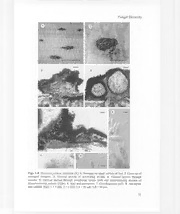
Figs. 1-8. Dianesea palmae, lectotype (K). 1.Stromata on upper surface of leaf. 2. Close-up of
stromatal complex. 3. Vertical section of developing stroma. 4. Vertical section through
ascoma. 5. Vertical section through conidiomaI locule (left) and accompanying ascoma of
Hysterostomina palmae (right). 6. Asci and ascospores. 7. Conidiogenous cells. 8. Ascospore
and conidia. Bars: 1= 5mm; 2 = 1mm; 3-4 = 50 /-lm;5-8 = 10/-lm.
73
-~-.l 9 10
1'j,'1f'1 -'((ln~;r>r)" ",.
,,~,~§
"US k'6Q:i'j1'(1i "
i
11
~II
~
'"
~ ~ '\0 'J
~
~ .'.~,,''~L·"",It~
~
~ ,--I-'l,l,-,(1 '1,;;/"\.,
nn·
I'.
nno,'" 1'1<,",~
~(lnl'(l, I, 0
iV}\.')I_,
~r;\, ;"'\'- .'rIn·rin"llcWr',\~!");"' r.;r' :'J"1"Y~/0-(2/pJ\ICO~J,\~'CJ 12 13
Figs. 9-13. Dianesea palmae, lectotype (K) and Hysterostomina palmae. 9-11. Dianesea palmae. 9. Vertical section through ascoma.
10. Detail of stromata and ascomatal wall. 11. Conidiogenous cells and conidia. 12. Vertical section through stroma (left) and
accompanying ascoma of Hysterostomina palmae (right). 13. Asci and ascospores of Dianesea palmae (left) and Hysterostomina
palmae (right). Bars: 9,12 = 100 IJ-m;]0 = 50 IJ-m;]],13 = 20 /Jm.
Fungal Diversity
Teleomorph: Habit appearing at the same time as, or possibly after the
conidiomata, often occupying the same stromatic crusts. External appearance
brown to dark-brown, perithecial, immersed in the stromatic crust, forming
irregular warts. In horizontal section with an upper wall composed of textura
angularis with cells 3-7 !lm diam with dark brown walls. In vertical section
213-325 !lm deep, composed of a brown to dark-brown outer wall enclosing a
central fertile locule, usually with a small portion of displaced cuticle and
epidermis above the stroma, and with a basal stromatic layer mixed with plant
cells. Stromatal wall dense, thick, making observation of individual layers
difficult, composed of brown-walled cells 3-8 !lm diam, ± globose around the
edge and angular and somewhat larger in the central part. Ascomatal wall
composed of several layers of compressed cells, sometimes intergrading with
but easily distinguishable from the stroma due to their darker colour. Locule
83-238 x 100-218 !lm, composed of a thin basal subhymenial cushion 18-30
!lm deep, with asci and paraphyses above. Interascal tissue composed of
cellular pseudoparaphyses, mostly colourless, septate, thin-walled, filiform but
rounded and slightly swollen at the tips, 1-2 !lm thick, sometimes
dichotomously branched, the branches arising at an acute angle from the
middle or near the top of individual cells. Asci maturing sequentially, with
young and old asci in the same locule. Young asci variable in shape before
spores can be distinguished, normally cylindrical or cylindric-clavate to
clavate, at first thin-walled, becoming thick-walled particularly in the upper
part, with a subapical chamber formed before spores are visible. Full-sized asci
containing spores, 54-72 x 12-16 !lm, cylindric-clavate to clavate, thick-walled
particularly in the upper part, not changing colour in iodine, with 6 or 8 spores
arranged in one, two rows or in a cluster. Asci after spore release collapsed,
with a large apical crack in the outer wall, and with the inner wall extending
like a tongue through the crack. Ascospores 12-20 x 3-5 !lm, initially
colourless, guttulate, becoming light brown to brown, covered by a gelatinous
layer, verrucose, I-septate, slightly narrowed at the septum, cylindric
ellipsoidal, the lower cell slightly attenuated and rounded towards the base, the
upper cell with a rounded apex.
Anamorph: coelomycetous. Habit intimately mixed with ascomata, often
occupying the same stromatic crusts. External appearance difficult to
distinguish from the teleomorph, brown to black, shiny, crustose, embedded
within a common stroma. Conidiomatal locule 118-238 x 80-188 !lm,
contaning conidia and with conidiogenous cells lining all of the internal wall
surface. Conidiogenous cells 6-12 x 2-4 !lm, ampulliform to ampulliform
cylindrical, colourless, smooth. Conidia: 11-17 x 2-4 !lm, colourless, aseptate,
smooth, variable in shape, cylindric-ellipsoidal, ovoid or cylindric-clavate,
75
guttulate. Conidial development by a replacement wall-building apex system
with enteroblastic percurrent non-progressive proliferation (replacement wall
building apex phialides).
Discussion
Hysterostomina palmae
The discoid fruit bodies in the specimen originally described as Cocconia
palmae are poorly preserved, but appear very similar to the fungus described as
Hysterostomina palmae (Stevens, 1923). Hysterostomina was originally
described as a counterpart of Hysterostomella which lacked interascal tissue,
but is now regarded as a synonym of that genus. Hysterostomella itself is
poorly defined and possibly polymorphic, and will be the subject of further
monographic treatment. At least two other species of Hysterostomella are
known from palms, H. sparsa (Peck & Clinton) Barr (syn. H. sabalicola Tracy
& Earle; Barr et al., 1986) and H. elaeicola Maubl. Both are quite distinct in
ascospore measurements.
Dianesea palmae
Stevens (1927) in his description of Cocconia palmae mentioned: "The
loculiferous stroma occurs either as the superficial stroma or on the erumpent
stroma. The circular arrangement of the perithecia is so irregular that the
suggestion of this as character is questionable. This fungus is of special interest
as a form showing the characters of the Microthyriaceae in the radiation of the
superficial stroma, and of the Dothideales in possessing globose locules in a
stroma". The difficulty Stevens had in assessing the relationships of his species
I I
can clearly be attributed to his failing to appreciate that two fungi were present
and that his description was therefore a composite. The International Code of
Botanical Nomenclature allows the name to be typified subsequently by one of
the original elements. We have therefore chosen the perithecial fungus to
represent the name Cocconia palmae as it is the most prominent of the two.
Stevens also described and illustrated the asci and ascospores of this species
rather than those of the Hysterostomina (Stevens, 1927).
Cocconia palmae as newly circumscribed is now well characterised, but
its relationships remain unclear. It clearly cannot be maintained as a species of
Cocconia, a member of the Parmulariaceae (Dothideomyetidae) with ascomata
which release their spores through irregular splits rather than the distinct
ostioles found in C. palmae. An alternative placement is problematic, and an
appropriate, previously existing genus could not be found. The familial
affinities of the new genus are also not clear, but it seems most closely allied to
76
Fungal Diversity
Table 1.Morphological characters of Coccoidea, Coccoidella and Dianesea.
CBLLCpCBUsoaolelrneaaocclckuclvkwucunkdaiollnon,aotaie,wrgdpr,,peaneearpfllalCALdCpCHcBsLliatepiseyaisocpaoeyrlcysshpetlaikcclsectlieaipuytlciitnoBDkuisvLPdPCpCfHcrcalunnuhdnskiviediyioleesteeltoslncdeneyiydynaog,epuaoearrfairrstsclIerilrsaagdpirirtiiuclilicini,cunepuatpalnlutneacakndah,iue-wstlerechtdllr-nd,eteeaopesoanlldacloeyraeralaaretcperiplilsia,slbcrcaaciirpacnlehetapkiwoav-r-taepesylteiosactital,hraaenhrsolepltrylelraetgeyipehlpodovs,wsbgaiytrmasaephlrueabsieletsosetsile,eohodawes,tbridnmrdnayrltoae,ioidlw,wns,deenlel,l,-
OItAAASinstsssstrstecccouiriooomeamslsepacatoaatrales spdsmeuisplotvtiaonitntcehattenweaalrlthe base, sfimssuiutulhnicate form
Anamorph
the Coccoideaceae. An appropriate genus could not be found in the extensive
list of ascomycetes known from palms (Hyde et al., 2000).
The Coccoideaceae contains a single biotrophic genus Coccoidea Henn.
with a possible further member Coccoidella Hohn. (Kirk et al., 2001). The
circumscription of the family is largely similar to that of Dianesea, but
ascomata are formed as multiple distinct locules in peltate stromata, and the
anamorph is acervular rather than locular (stromatic). Coccoidea also has
ascospores which are septate close to the ends in contrast to those of Dianesea
where they have a ± median septum (Eriksson, 1981; Barr, 1987; Sivanesan,
1987; Kirk et al., 2001). Coccoidella forms irregular or botryose stromata
(Sivanesan, 1987); it is currently considered to be of uncertain familial
placement, but shares many characters with the Coccoideaceae (Kirk et al.,
2001). The three genera are compared in Table 1. Potentially the most
fundamental distinction between Dianesea and the genera of the
Coccoideaceae is the apparently ascohymenial type of ascomata with distinct
walls, rather than the ascolocular type which is typical of the
Dothideomycetidae. This might invite placement of Dianesea in the
Sordariomycetidae, but fissitunicate asci with rostrate dehiscence are unknown
in that group.
No other potential family placement within the Dothideomycetidae
appears appropriate. The Venturiaceae are mostly necrotrophs, but a small
proportion exhibit biotrophic nutrition. The family is currently poorly defined,
but most genera have small setose ascomata and hyphomycetous anamorphs, in
contrast to Dianesea (von Arx and Muller, 1975; Sivanesan, 1984; Barr, 1987;
77
I -I
Kirk et al., 2001). The genus Rosenscheldiella has some resemblance to
Dianesea with its external stromatal shape, but these contain multiple
ascomatal locules producing colourless ascospores but without a distinct
periphysate ostiole (Barr, 1987). The Botryosphaeriaceae contain a few
biotrophic species, but typically lack interascal tissue and have aseptate
ascospores. The Mycosphaerellaceae are primarily necrotrophs and saprobes
and rarely have any significant level of stromatic development, but some
species of Microcyclus are biotrophic and appear superficially similar to
Dianesea. However, they lack interascal tissue (Cannon et al., 1995). The
Dacampiaceae show some similarities with Dianesea, but stromatic tissue is
poorly developed, ascomatal walls are much thicker and multilayered, and
many are lichenicolous or lichenised (Barr, 1987). The Parodiellaceae contains
biotrophic fungi, but have well-developed verrucose unilocular ascostromata.
The Phaeosphaeriaceae have thin-walled ascomata which are occasionally
aggregated into multilocular stromata, but are saprobes or necrotrophs and
have quite distinct anamorphs.
Acknowledgements
Research access to the herbarium of the Royal Botanic Gardens, Kew and assistance
from B. Spooner is gratefully acknowledged. The advice and informatics support of D. Minter
(CAB! Bioscience) played a major role in establishing the research programme. The senior
author thanks CNPq (Conselho Nacional de Desenvolvimento Cientifico e TecnoI6gico),
Brazil for financial support.
References
Arx, l.A. von and MUlier, E. (1975). A re-evaluation of the bitunicate Ascomycetes with keys
to families and genera. Studies inMycology 9: 1-159.
Barr, M.E. (1987). Prodromus to the class Loculoascomycetes. Published by the author, USA.
Barr, ME, Rogerson, C,T., Smith, S.J. and Haines, lH (19Rfi). An annotated catalog of the
pyrenomycetes described by Charles H. Peck. Bulletin of the New York State Museum
459: 1-74.
Cannon, P.F., Carmanin, c.c. and Romero, A.!. (1995). Studies on biotrophic fungi from
Argentina: Microcyclus porlieriae, with a key to South American species of
Microcyclus. Mycological Research 99: 353-356.
Eriksson, O.E. (1981). The families ofbitunicate ascomycetes. Opera Botanica 60: 1-210.
Hyde, K.D., Taylor, lE. and Frohlich, l (2000). Genera of Ascomycetes from Palms. [Fungal
Diversity Research Series I], Fungal Diversity Press, Hong Kong.
Kirk, P.M., Cannon, P.F., David, l.C. and Stalpers, l.A. (2001). Ainsworth & Bisby's
Dictionary of the Fungi. (9th ed.). CAB! Publishing Wallingford, axon, UK.
Sivanesan, A. (1984). The Bitunicate Ascomycetes and their Anamorphs. Vaduz, l.Cramer.
Sivanesan, A. (1987). Coccoidella perseae sp. novo and its anamorph Colletogloeum perseae
sp. novoTransactions of the British Mycological Society 89: 265-270.
78
Fungal Diversity
Stevens, F.L. (1923, pub!. 1924). Parasitic fungi from British Guiana and Trinidad. Illinois
Biological Monographs 8: 173-242.
Stevens, F.L. (1927). Fungi from Costa Rica and Panama. Illinois Biological Monographs 11:
159-254.
(Received 23 December 2001; accepted 20 January 2002)
79

